What does a normal or abnormal treadmill test mean?

TREADMILL testing is a common modality used by many doctors to screen for heart artery disease. For the couch potatoes who are suddenly fired up to become a paragon of fitness, current guidelines recommend that it will be wise for relatively inactive older men to undergo a treadmill stress test before embarking on a vigorous exercise regime. If the treadmill test result appears normal, does that mean that you do not have heart artery disease and you will not get a heart attack? If the test result is abnormal, does it mean that you have significant heart artery disease?

What is a treadmill stress test?

The treadmill stress test, though a commonly used screening test by doctors, has many limitations, especially for women. Patients are hooked up to a monitor which records the electrical pattern of the heart – also known as an electrocardiogram (ECG) – and walk on a treadmill machine which increases in speed and incline every few minutes according to the preset protocol. During the test, doctors look for changes in the ECG to screen for heart artery disease.

Treadmill test in women

Research from the last decade has revealed gender differences in heart disease. American Heart Association statistics have shown that 64 per cent of women who died suddenly from heart disease did not have classic warning symptoms, versus 50 per cent of men.

Moreover, 38 per cent of women died within one year following a heart attack, versus 25 per cent of men. Hence, there is a need to detect underlying heart artery disease in women. Although treadmill testing is widely used, the pitfalls of this method need to be understood.

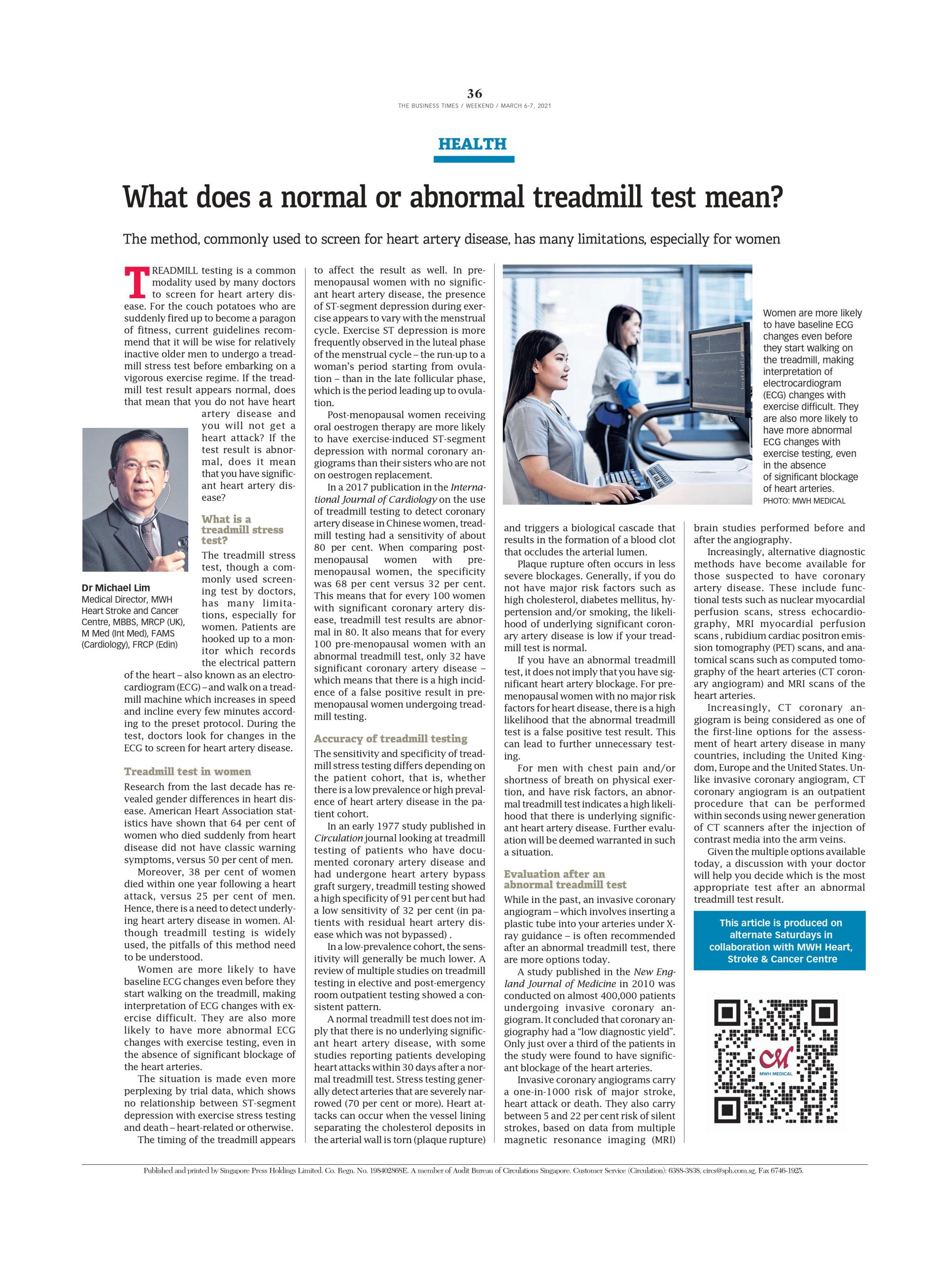
Women are more likely to have baseline ECG changes even before they start walking on the treadmill, making interpretation of ECG changes with exercise difficult. They are also more likely to have more abnormal ECG changes with exercise testing, even in the absence of significant blockage of the heart arteries.

The situation is made even more perplexing by trial data, which shows no relationship between ST-segment depression with exercise stress testing and death – heart-related or otherwise.

The timing of the treadmill appears to affect the result as well. In pre-menopausal women with no significant heart artery disease, the presence of ST-segment depression during exercise appears to vary with the menstrual cycle. Exercise ST depression is more frequently observed in the luteal phase of the menstrual cycle – the run-up to a woman’s period starting from ovulation – than in the late follicular phase, which is the period leading up to ovulation.

Post-menopausal women receiving oral oestrogen therapy are more likely to have exercise-induced ST-segment depression with normal coronary angiograms than their sisters who are not on oestrogen replacement.

In a 2017 publication in the International Journal of Cardiology on the use of treadmill testing to detect coronary artery disease in Chinese women, treadmill testing had a sensitivity of about 80 per cent. When comparing post-menopausal women with pre-menopausal women, the specificity was 68 per cent versus 32 per cent. This means that for every 100 women with significant coronary artery disease, treadmill test results are abnormal in 80. It also means that for every 100 pre-menopausal women with an abnormal treadmill test, only 32 have significant coronary artery disease – which means that there is a high incidence of a false positive result in pre-menopausal women undergoing treadmill testing.

Accuracy of treadmill testing

The sensitivity and specificity of treadmill stress testing differs depending on the patient cohort, that is, whether there is a low prevalence or high prevalence of heart artery disease in the patient cohort.

In an early 1977 study published in Circulation journal looking at treadmill testing of patients who have documented coronary artery disease and had undergone heart artery bypass graft surgery, treadmill testing showed a high specificity of 91 per cent but had a low sensitivity of 32 per cent (in patients with residual heart artery disease which was not bypassed) .

In a low-prevalence cohort, the sensitivity will generally be much lower. A review of multiple studies on treadmill testing in elective and post-emergency room outpatient testing showed a consistent pattern.

A normal treadmill test does not imply that there is no underlying significant heart artery disease, with some studies reporting patients developing heart attacks within 30 days after a normal treadmill test. Stress testing generally detect arteries that are severely narrowed (70 per cent or more). Heart attacks can occur when the vessel lining separating the cholesterol deposits in the arterial wall is torn (plaque rupture) and triggers a biological cascade that results in the formation of a blood clot that occludes the arterial lumen.

Plaque rupture often occurs in less severe blockages. Generally, if you do not have major risk factors such as high cholesterol, diabetes mellitus, hypertension and/or smoking, the likelihood of underlying significant coronary artery disease is low if your treadmill test is normal.

If you have an abnormal treadmill test, it does not imply that you have significant heart artery blockage. For pre-menopausal women with no major risk factors for heart disease, there is a high likelihood that the abnormal treadmill test is a false positive test result. This can lead to further unnecessary testing.

For men with chest pain and/or shortness of breath on physical exertion, and have risk factors, an abnormal treadmill test indicates a high likelihood that there is underlying significant heart artery disease. Further evaluation will be deemed warranted in such a situation.

Evaluation after an abnormal treadmill test

While in the past, an invasive coronary angiogram – which involves inserting a plastic tube into your arteries under X-ray guidance – is often recommended after an abnormal treadmill test, there are more options today.

A study published in the New England Journal of Medicine in 2010 was conducted on almost 400,000 patients undergoing invasive coronary angiogram. It concluded that coronary angiography had a “low diagnostic yield”. Only just over a third of the patients in the study were found to have significant blockage of the heart arteries.

Invasive coronary angiograms carry a one-in-1000 risk of major stroke, heart attack or death. They also carry between 5 and 22 per cent risk of silent strokes, based on data from multiple magnetic resonance imaging (MRI) brain studies performed before and after the angiography.

Increasingly, alternative diagnostic methods have become available for those suspected to have coronary artery disease. These include functional tests such as nuclear myocardial perfusion scans, stress echocardiography, MRI myocardial perfusion scans , rubidium cardiac positron emission tomography (PET) scans, and anatomical scans such as computed tomography of the heart arteries (CT coronary angiogram) and MRI scans of the heart arteries.

Increasingly, CT coronary angiogram is being considered as one of the first-line options for the assessment of heart artery disease in many countries, including the United Kingdom, Europe and the United States. Unlike invasive coronary angiogram, CT coronary angiogram is an outpatient procedure that can be performed within seconds using newer generation of CT scanners after the injection of contrast media into the arm veins.

Given the multiple options available today, a discussion with your doctor will help you decide which is the most appropriate test after an abnormal treadmill test result.

When is sex too stressful for your heart?

WHILE having sex is a normal part of life, many wonder whether their heart can continue to withstand the excitement of sex when they grow older. Those with heart disease worry about the risk of precipitating a heart attack during sex.

Many with heart disease have associated risk factors such as diabetes mellitus, hypertension, smoking and medication, which may impair the ability to engage in sex. While the use of drugs such as Viagra may alleviate the situation, a common concern is the impact of such drugs on those with heart disease.

Sex as exercise

Sex does burn calories. With sexual arousal and physical exertion, adrenaline increases and the heart rate and blood-pressure increases. Most studies have shown that, despite the perception of sex being a vigorous physical activity, the heart rate generally remains below 130 beats a minute, and systolic blood pressure below 170, while having sex with a familiar partner.

On the average, middle-aged couples have sex about twice a week, each episode lasting about 15 minutes, and burning more than 50 to 80 calories depending on the intensity of sex.

Activities that will burn about 50 calories will include running up and down the stairs for three minutes or playing tennis for six minutes.

Will sex increase the risk of heart disease or stroke?

Though some people get headache during sex and worry about getting a stroke, a study published in the Journal of Epidemiology and Community Health found that frequency of sex was not associated with stroke in the 914 men they followed for 20 years.

A 10-year study on the sex habits of about 2,400 men in a town in Wales showed that those who reported the most frequent sexual activity were only half as likely to suffer a stroke or heart attack during that time. Hence, from an epidemiological perspective, sex does not increase the long-term risk of heart attack or stroke.

This could be partly explained by the reduction of stress and blood pressure with sexual activity. Studies reported in the journal Biological Psychology showed that cohabiting participants who had frequent sexual intercourse had better response to stress and lower diastolic blood pressure.

Sexual activity and orgasms increase the levels of the hormone oxytocin (also termed the “love hormone”).

Higher levels of oxytocin are associated with the urge to bond and with the feeling of generosity. As the level of oxytocin hormone increases, endorphins also increase. Endorphins have morphine-like effects on the brain and hence the sensation of pain declines. The oxytocin released during orgasm also promotes sleep.

Hence, you should not be surprised as to why your partner falls asleep so easily after sex. There is substantial data that getting adequate sleep is associated with a lower risk of heart disease and stroke.

Can sex cause a heart attack?

In a study published in the Journal of the American Medical Association, researchers from Tufts Medical Centre in Boston concluded that exercise and sex can increase the risk of heart attack and sudden cardiac death, although the increased risk is small and transient.

The risk is higher for those who exercise infrequently compared to those who exercise regularly. The researchers pooled together smaller studies that compared the risk of heart attacks and sudden death when participants were exposed to physical activity and engaged in sexual activity to times when they were not.

The study found that episodes of physical activity increased heart attack risk 3.5 times and risk of sudden cardiac death nearly five times.

Sexual activity increased heart attack risk 2.7 times but there was no data available on the link between sex and sudden cardiac death risk. The relative risk is a comparison of the risk when participants are exercising or having sex compared to the risk when they are not.

What it means is that physical exercise and sexual activity can trigger the onset of heart attack and sudden cardiac death, but that risk is transient and lasts for the one to two hours during and after the activity. While the relative risk of a heart attack during sex is 2.7 times, the absolute risk (that is, the probability of heart attack or sudden cardiac death actually occurring in an individual) is extremely small, at least during conventional sex with a familiar partner.

To look at it from another perspective, for a healthy 50-year-old man, the risk of having a heart attack in any given hour is about one in a million. Sexual activity doubles the risk, but even then, the risk is minuscule – just two in a million.

A study published in Lancet by Belgian researchers reported that 2.2 per cent of heart attacks is related to sexual activity. The million-dollar question for many men with heart disease is: “What is the risk of getting a heart attack during sex?” Yes, the risk is increased by 10 times compared to healthy men but even with this increased risk, the likelihood of getting a heart attack during sex is only one in 50,000.

A study on sudden cardiac arrest during sexual intercourse published in the Circulation journal in 2018 examined the data from the Paris-SDEC registry (Paris-Sudden Death Expertise Center). For those who had sudden cardiac arrest and were admitted alive to the hospital, less than one per cent were sex-related and occurred typically among middle-aged men with cardiovascular risk. About seven in 10 were due to underlying heart disease and about three in 10 were due to subarachnoid bleeding (bleeding between the skull and the brain), a type of intracranial bleeding due to rupture of an aneurysm of a brain artery.

ABCs of having sex safely

Most people with heart conditions want to know when they should avoid sex, and when they can begin having sex again. Avoid sex if you are unwell or if you detect symptoms of heart disease such as shortness of breath or chest tightness on exertion. If these symptoms occur during sex, stop immediately.

Not uncommonly, doctors are confronted with the question: “Is it safe for me to take Viagra?” Common drugs for males with erectile dysfunction (resulting in inability to complete sexual intercourse) include sildenafil (Viagra), vardenafil (Levitra) and tadalafil (Cialis). For those with heart disease, being able to exercise up to a heart rate of 130 per minute without any heart symptoms will generally indicate that it is safe to have sex, even with the use of such medication. There is one very important contra-indication – those who take nitrate medications cannot take these drugs.

Simply put, if you feel well after walking up the stairs for at least three floors, you can safely assume that your heart can withstand the “stress” of sex.

The highs and lows of blood pressure in older adults

BEYOND the wrinkles and the blemishes that come with advancing years, the invariable march of age will result in changes in the blood vessels that have served us so faithfully. Just as rubber becomes stiffer and less pliable over time, our arteries will also lose its elasticity and become less distensible. An effect of this is an increase in the upper or systolic blood pressure (SBP) value and labile fluctuation in SBP with varying levels of activities and emotional changes.

Hence, it is not uncommon to find in the elderly a blood pressure (BP) of 160/90 mm Hg at the doctor’s clinic, even though the preceding home BP recording showed 130/80 mm Hg.

“High” blood pressure that is normal

One elevated blood pressure reading does not mean that high blood pressure (hypertension) exists. While the pliable and elastic wall of the arteries in a young person allows the arteries to distend and “absorb” the increase pressure that is generated through an increase in heart rate or by stress, the loss in elasticity and stiffening of arteries with ageing means an elevation in the SBP will be more commonly seen with emotional stress, anxiety and physical exercise. Hence, in the elderly, the need to record blood pressure on multiple occasions at rest is necessary before a diagnosis of hypertension can be confirmed.

However, office and home blood pressure measurements may still be elevated in the situation of “pseudo-hypertension”, a condition where the SBP is falsely elevated because of significant stiffening and hardening of the walls of the arteries. In this situation, the cuff inflation during blood pressure recording may have to be inflated to very high pressures before the artery can be compressed, resulting in a highly elevated SBP value and a normal lower or diastolic blood pressure (DBP) value.

This may be more often seen in those who have diabetes mellitus where diffuse hardening of the arterial wall is more commonly seen. Furthermore, smaller arteries are more likely to be affected as compared to larger arteries.

Hence, in this situation, it may be better to use an arm BP measuring device rather than a wrist BP measuring device. This condition should be suspected if there are absolutely no symptoms and no evidence of any damage to any organs, despite the BP being persistently high and not responding to medication.

The three situations of labile SBP elevation, “white-coat hypertension” and “pseudo-hypertension” are more prevalent in the elderly and a correct diagnosis is important in avoiding overtreatment.

Impact of high blood pressure

Persistent elevation of high BP can manifest itself by the presence of damage to the brain (stroke, bleeding, dementia), the heart (chamber enlargement, abnormal heart rhythms), major blood vessels (aneurysm or enlargement of the aorta), kidneys (impairment of function) and eyes (degeneration of eyesight).

An objective way to assess the effect of persistent hypertension is to evaluate the impact of high BP on the heart. The left-side heart chambers are connected to the aorta, the main arterial channel which supplies blood to the entire body. In persistent poorly controlled hypertension, the thinned walled left upper heart chamber will be enlarged and the muscular left lower chamber will have thickened walls as the heart chamber has to pump harder against a higher BP for the blood to enter the aorta. This can be objectively confirmed by an ultrasound examination of the heart, which is also called echocardiography.

The absence of these changes on echocardiography in the presence of persistently high BP not responsive to medication and yet not accompanied by symptoms should lead one to suspect “pseudo-hypertension”.

How low should the blood pressure be?

The latest American College of Cardiology and the American Heart Association guidelines recommend that older adults with a BP of 130/80 mm Hg or higher should be treated. Under normal resting conditions, about 85 per cent of the blood perfusion of the muscle of the main heart pumping chamber, the left ventricle, occurs during diastole (relaxation phase of the heart chamber). Hence, the arterial diastolic BP (DBP) is the main force that “pushes” blood into the heart arteries to perfuse the left ventricle muscle.

Presently, we do not know what is the lowest limit of DBP where it is not able to maintain adequate perfusion to the left ventricle muscle. In patients with heart artery disease, if the cross-sectional area of the heart artery is reduced by more than 70 per cent , there will usually still be adequate perfusion due to compensatory dilatation of the microcirculation but the maximum heart artery blood flow will be reduced.

If the cross-sectional area reduction is at least 85 per cent to 90 per cent, there can be inadequate perfusion even at rest. Hence, in those with significant blockage of the heart arteries, the DBP should not be excessively low and should be individually titrated. Likewise, in those with significant narrowing of the brain arteries, a sudden drop in the SBP can precipitate a stroke. Hence, for the elderly diabetics who are at a higher risk of developing heart and brain artery disease, caution should be exercised.

In the ACCORD BP trial that looked at intensive BP lowering in diabetics, intensive BP control (SBP <120 mmHg) did not reduce the combined major cardiovascular complications (heart attack, stroke and death). However, when the outcomes were looked at individually, there was a reduction in stroke at the expense of increased serious adverse events. Hence, while achieving BP targets <140/90 mmHg may be suitable for some diabetics, not all will be better off with intensive BP lowering (SBP <120 mmHg).

In the International Database on Home Blood Pressure in Relation to Cardiovascular Outcome (IDHOCO) study published in Circulation journal in 2015, for those hypertensive octogenarians treated, a SBP of less than 127 mm Hg was associated with increased death and the lowest risk was at a systolic BP of about 149 mm Hg. Hence, for hypertensive octogenarians, as a result of age-related decreased circulation to the organs and a higher risk of a postural drop in BP on standing, maintaining the SBP between 150 mmHg and 140 mmHg is the sweet spot.

Should I worry about the pain in my chest?

Should you worry about the chest discomfort you have been having lately? Every so often, when you are jogging, playing your game of tennis or while you are enjoying your game of golf, you may have felt that discomfort in the chest and wondered whether to dismiss it or take it as a warning of underlying heart disease. This is a common dilemma.

Worrisome chest discomfort

Chest discomfort or pain is the most common complaint encountered by family physicians, cardiologists, and emergency-room physicians. What distinguishes the chest pain due to underlying blockage of the heart arteries (angina pectoris) from other types of chest pain are the characteristics of the pain. The term angina pectoris is derived from the Latin word angina (“infection of the throat”), the Greek ankhone (“strangling”), and the Latin pectus (“chest”), and can therefore be translated as “a strangling feeling in the chest”.

Hence, during exercise, the increased demand for oxygen is met primarily through increased blood flow through the heart arteries. If there is an obstruction to blood flow because of the narrowing of heart arteries, the oxygen supply may be unable to meet the increased demand, resulting in an environment where there is insufficient oxygen. This results in the activation of cellular pathways which operate in an oxygen-scarce environment, resulting in the production of chemicals such as lactic acid. The build-up of these chemicals stimulates nerve endings that cause the sensation of pain.

Angina or heartburn?

Heartburn is an uncomfortable feeling of burning or warmth in the central chest, which may radiate to the neck, throat, and jaw. It can mimic angina and present itself as chest tightness. Unlike angina, it is not due to heart disease, but is a result of backflow of acid from the stomach into the oesophagus (gastroesophageal reflux disease or GERD). It is typically aggravated by lying down or bending over soon after a meal and relieved by standing up, drinking water, and taking antacids. It is present in about one-third of adults, especially in pregnant women. It can sometimes be difficult to distinguish it from angina, the main distinguishing factor being that heartburn is not related to exertion.

Angina in the young

If you are young and have chest tightness or shortness of breath while running and never seem to be able to keep up with others during exercise, it may not be that you are not physically fit. There is also a possibility that you may have underlying congenital heart artery disease. Angina can occur in the young as a result of inherited conditions. For the majority, while the major heart arteries lie on the surface of the heart, in some, the artery may take a course where a segment of the artery may be embedded in the heart muscle (myocardial bridging) and hence the embedded segment may be compressed by the heart muscle bands during contraction of the heart.

If a long segment is embedded deeply into the heart muscle, the obstruction to flow may be significant enough to cause angina during vigorous physical exertion. Another rare cause of mechanical obstruction is the abnormal origin and course of the heart artery, where a segment of it is wedged between the main artery arising from the heart (aorta) and the lung artery (pulmonary artery). During heavy physical exertion, the pulsations of the two large arteries may result in compression of the heart artery to the extent that it causes angina. On rare occasions, these two inherited causes of heart artery obstruction can cause sudden death during vigorous physical exertion in the young.

Angina not related to exertion

While angina is typically associated with physical exertion, there is an uncommon variety called coronary vasospasm or Prinzmetal’s angina. This condition is due to transient constriction of the heart artery secondary to abnormalities in the regulation of the smooth muscle in the wall of the artery. The angina associated with spontaneous spasm of the heart artery usually develops during sleep or early in the morning. In some patients, chest pain development is associated with alcohol drinking and one interesting feature is that the chest pain episode tends to develop several hours after alcohol intake, rather than occurring immediately after drinking. Hence, these patients may be mistakenly diagnosed to have gastrointestinal problems including stomach ulcer disease or GERD. Coronary vasospasm, if unrecognised, can result in heart attacks and sudden cardiac death.

Chest pain but not angina

Understanding angina will help to distinguish the types of chest pain that do not need urgent medical attention. Chest pain is usually not angina if it can be localised to a single site on the chest using two fingers, is sharp, pulling or pinprick in nature, and not aggravated by exertion. If you are young and cannot seem to be able keep up with your friends when running, the presence of chest pain during running may suggest that you may have an underlying inherited heart artery condition. Medical advice should be sought before embarking on vigorous endurance exercise activities such as marathons. If you have multiple risk factors for heart disease and have typical exertional anginal chest pain, it is very likely that you have underlying blockage of your heart arteries. You should have your heart assessed by your doctor before attempting vigorous physical activities.

However, if you have risk factors for heart disease and have no symptoms, do remember that having no chest pain does not mean there is no heart disease. In a US study on out-of-hospital sudden cardiac arrest published in the Circulation journal in 2006, only about 25 per cent had angina prior to the sudden collapse. In that study, only 6 per cent survived the sudden cardiac arrest and were discharged from the hospital alive. Hence, controlling your risk factors and having regular follow-up with your doctor is the best way to prevent sudden death.

Life extension – health, rejuvenation and longevity

The relentless pursuit of the elixir of youth has spurred anti-ageing research in attempts to achieve the triple goals of life extension, namely, the triumvirate of healthy lifespan, rejuvenation and longevity. Sirtuins are a family of cellular enzymes that are powered by a chemical compound called nicotinamide adenine dinucleotide (NAD). They play an important role in preventing diseases and even reversing some aspects of ageing. Studies have shown that increased sirtuin activity in mammals has been associated with a delayed onset of age related diseases and increased longevity.

Increased sirtuin activity appears to inhibit nerve degeneration and reduces the development of cardiovascular and metabolic diseases (such as diabetes mellitus and abnormal lipid levels). Hence, if sirtuin activity can be increased using compounds that can boost its activity (STACs or Sirtuin Activating Compounds), the use of STACs can potentially help a person stay healthy longer, even if longevity is not affected.

Resveratrol
Resveratrol is a natural plant phenol STAC found in the skin of red grapes and other fruits such as blueberries and cranberries. Contrary to common belief, red wine contains very little of it. Resveratrol has been shown to have life-extending properties in studies on lower-order species such as yeast and nematodes, but this effect has marginal reliability in higher-order species. Nevertheless, it has been shown to have potentially beneficial effects. Before you start taking large doses of resveratrol, you may be surprised to know that it is a Janus-faced compound.

Low dietary doses may suffice to elicit the biological responses required to optimise the body’s defence mechanisms against incipient disease. But at high doses, it behaves in a contrarian manner. At low doses, resveratrol induces responses that overlap with the female hormone oestradiol. Low-dose effects seen in animal and human studies include beneficial metabolic effects such as more efficient glucose reduction in diabetics, reducing the development of obesity and non-alcoholic fatty liver disease, protecting against arterial degeneration, delaying development of neurodegeneration, and improving motor and cognitive functions.

At high doses, resveratrol has an anti-oestrogen effect which suggests that it may reduce the risk of oestrogen- dependent cancers. This Janusfaced hormetic effects of resveratrol may partly explain the French paradox, where there is a reduced incidence of cardiovascular diseases and certain types of cancer in some populations despite their consumption of high-fat diets.

NAD
NAD is an important molecule that is essential for over 500 enzyme reactions in the body which impact metabolism, ageing, cell death, DNA repair, and gene expression. Hence, NAD plays a pivotal role in human health span and longevity and is a necessary substrate for sirtuin enzymatic activity. In mammalian cells, NAD is mainly generated by the conversion of nicotinamide (a soluble form of Vitamin B3) into nicotinamide mononucleotide (NMN) followed by its combination with another molecule to form NAD. There is also another precursor, nicotinamide riboside (NR), that is converted by enzymes to NMN in the cells. As this pathway is safe and also the most efficient route for production of NAD, NMN or NR supplementation has been used to increase NAD levels.

Animal studies have shown that NMN supplementation can ameliorate the age-related reduction in NAD production in cells and improve the body’s cardiovascular response to ageing. Age-related decrease in arterial elasticity means that the aorta is less able to expand and buffer the increased blood pressure generated each time the heart pumps. With ageing, the production of a pressure-bearing protein, type 1 collagen, in the arterial wall increases, whereas the main protein responsible for the structural integrity and elastic properties of the arterial wall, elastin, decreases. Studies in mice have shown that NMN supplementation was able to reduce stiffness in large arteries by reversing the accumulation of type 1 collagen in arterial walls and improving elastin content.

From the age of 40 years onwards, there is a gradual decline in perfusion of the body tissues resulting in gradual deterioration in body function towards the last decades of life. A consequence of this is cognitive decline. Optimal brain function is dependent on adequate oxygen and nutrient delivery via minute brain blood vessels (cerebral microvascular circulation). This modulation of brain blood flow in response to increased brain activity is impaired with age, contributing to age related cognitive impairment. Studies in aged mice given NMN have demonstrated an improvement in the modulation of cerebral microvascular circulation. Animal studies have also shown that NMN can prevent age-related cognitive decline by reducing cell death in areas of the brain that control short and long-term memory.

Use of NMN was also associated with decrease in the neurodegenerative changes seen in Alzheimer’s disease and age-related retinal changes. This age-related decrease in the production of new vessels and a gradual decrease of blood vessels in the microcirculation also result in reduction in muscle mass and diminishing exercise capacity with age. Mice given NMN were able to demonstrate an increase in the production of new vessels in the muscle and an increase in density of small vessels, thereby improving exercise capacity.

Caloric restriction
Caloric restriction (CR) which involves calorie reduction without causing malnutrition, has been associated with an increase in lifespan in some animal studies. In these studies, dietary CR was associated with increased lifespan and reduced disease incidence,  specially cancers. However, some studies did not show benefit and, in some mouse strains, CR was associated with shortened lifespan. Observational studies on humans who have practised extreme CR over many years showed low levels of risk factors for cardiovascular disease and diabetes. Similarly, a human study on CR, the CALERIE study, found that CR participants had lower blood pressure and lower cholesterol. The study was too short to examine the impact of CR on lifespan. The current conclusion from the National Institute of Aging in the United States is that there is not enough evidence to recommend CR as a therapeutic measure for life extension.

Although CR was associated with lower risk factors for cardiovascular diseases and diabetes, caution is urged as in a study using mouse lemurs on prolonged CR, MRI studies showed that there was more widespread age-related grey matter atrophy in CR animals while only a few regions in the brain showed atrophy in those not on CR.

Life extension
Globally, heart disease and stroke are the two main causes of death in most high-middle and high-income countries. Hence, the first cardinal principle in life extension is to control the risk factors such as blood pressure elevation, cholesterol elevation, sugar elevation and smoking. The second principle is to have a healthy lifestyle such as keeping the weight within the healthy range and exercising regularly. Both physical exercise and dietary CR result in a significant increase in NAD production and increase sirtuin activity. CR may be an option as part of a weight-reduction regime to keep the weight optimal. The third principle is to see your doctor regularly to control risk factors.

Finally, among the supplement options, scientific studies favour the use of low-dose resveratrol or NMN as they may potentially provide many health benefits via increased sirtuin activity, although more studies will be required to understand their efficacy in human life extension.

Glycemic Variability holds the key to diabetic complications

A FORTNIGHT ago, I wrote about my Apple watch that was a birthday present from my better half. We foresaw that the Apple watch would eventually be a telemetry device for human beings. One of the interesting upcoming developments by Apple is the ability to measure sugar non-invasively.

So what is it about sugar? In the ’60s and ’70s we demonised fat as the cause of heart attacks and strokes. From the last decade, we realised that in substituting sugar in the place of fats, we had inadvertently triggered an avalanche of obesity, diabetes and chronic diseases.

The pendulum had swung so much that major food companies globally had pushed the presence of sugar and corn syrup in processed foods, low fat products and fast foods. As the global population consumed more refined sugars, the propen-sity to have the sugar rush and the yearning for more sugar had triggered the medical equivalent of horsemen of the apocalypse.

So really, what is it about sugar? We know that there are ways to measure foods and their sugar content. The first is the Glycemic Index or GI. This essentially measures how fast the food turns to sugar in our gut and is absorbed into the blood stream. We assign a perfect score of 100 to a glass of sugar syrup. White rice is about 73, white bread is 75 while potato is in the 80s. Pasta, glass noodles, soba, udon and rolled oats are a lot better weighing in at the 50 range. You are obviously better off eating low GI foods.

The third term we should know about sugar is Glycemic Response or GR. This is essentially the beautiful peaks and troughs of our sugar levels related to our mealtimes. We found from the analytics that the peaks can be measured statistically in many ways. We can measure how fast the sugar peaks after a meal over time, we can measure the area under the curve of the peak, we can measure how fast the sugar drops from the peak over time and we can also measure the troughs. The troughs are interesting as some of the subjects have very low blood sugars after a huge peak. They are on the verge of hypoglycemia but have no symptoms or have no recollection, as they are fast asleep.

We know that there is something called “reactive hypoglycemia”. This is when one takes a big meal of high GI carbohydrates and there is a flood of insulin released. The insulin clears the sugar from the bloodstream but due to reasons unknown, the insulin surge and its after-effects seem to carry on to push the blood sugar to very low levels.

What is the point of measuring all these intricacies of the peaks and troughs of blood sugar? We then come to the fourth and final term called Glycemic Variability or GV. In measuring all these peaks and troughs related to food, we realise that a gentle pattern like a sine wave is much preferred to a jagged saw tooth pattern with huge swings.

There is now realisation that GV holds the key to all the dreaded complications we see in diabetics from the eye problems leading to blindness, to the deadened nerve endings that result in gangrene and amputations and to the kidney disease that ends in dialysis. We have also seen the hidden hand of GV in increased rates of heart attacks, strokes, hospitalisation and mortality. There is also suggestion that it may impact cognitive function, predict depression and accelerate formation of cancers.

Continuous glucose monitoring

With the advent of continuous glucose monitoring in the last few years, we have started looking at the GV with more granularity. Abbott is taking the continuous glucose monitoring system to the next level with its latest system recently announced in September, that will automatically deliver continuous real-time glucose readings to a user’s smartphone every minute. This is a major enhancement from its current system of measuring blood sugars every 15 minutes. Previously we could only measure a three-month average of blood sugars or ask the diabetic patient to prick himself seven times daily to give us an idea of how his blood sugars varied throughout the day.

So why is GV such a potent force? The jury is still out but there are some who think it mirrors the release of insulin and insulin triggers fat deposition in our bellies or what is called visceral fat. The visceral fat is different from fat on your thighs and buttocks. This visceral fat is like an “alien” organ. It releases all kinds of bad hormones and cytokines that increase the risk of heart attack, strokes and even cancer.

Can we then do something about the GV? The answer is obvious: if you know what kinds of meals spike your sugar or if you go hypoglycemic at night, you can change your diet and cut out foods that trigger the peaks and eat in a way to nullify the troughs. The way we respond to certain foods is quite individual and the GR may be different for different individuals and different foods; it is like you have your own individual pattern. Until you put on a glucose monitor, you cannot really tell.

The real benefit ultimately is to smoothen out your peaks and troughs with a combination of judicious and timed food intake, appropriate exercise and sound sleep, and the kilograms should come off as well as the centimetres off your waistline as you shed visceral fat. After all, all of us wish for a better quality of life and that quality reflects how we feel, move and look.

The Smart Watch: Telemetry For Humans?

GOOD nutrition, clean environments, accessibility to good healthcare and healthy lifestyles have increased human longevity. Nevertheless, the quest of increasing longevity is insatiable. As medical science pushes the boundaries of human lifespan, delving into the science of longevity can help us sieve through the reams of information and distil the essence of anti-ageing and cell preservation.

Cellular ageing

Cell ageing or cellular senescence is a physiological state with permanent cessation of the ability of the cell to replicate and accumulation of damaged DNA ( basic building blocks of genes), and this process is increased by ageing. This cellular senescence is marked by shortening of the protective terminal ends of the chromosomes which are called telomeres. Telomeres are like plastic caps at the ends of shoelaces, preventing them from being frayed and damaged. Shortening of the telomere is a sign of cellular senescence. In 1979, the first of the SIR (silent information regulator) proteins or sirtuins was reported. Over time more members of this protein group were found. In 1999, the publication of a study that reported that increased sirtuin activity could increase the lifespan of yeast cells by 70 per cent heralded a major breakthrough that accelerated the interest and research in sirtuins. Since then, there has been a growing body of evidence that the cell has a group of anti-ageing proteins, the sirtuins, that are essential for delaying cellular senescence and increasing the lifespan of organisms.

Anti-ageing proteins

Core to the science of anti-ageing is this group of proteins called sirtuins. In human cells, there are seven sirtuins (SIRT 1 to 7) with various roles within the cell. Three of these sirtuins (SIRT 3,4,5) control the generation of power for the needs of the cells and function in the mitochondria (power generator which produce the energy for the needs of the cell) with roles in cellular antioxidant balance and lipid metabolism. Another three sirtuins (SIRT 1, 6, 7) control the genetic framework in the cell and function in the cell nucleus (the central core of the genetic material in the cell) with roles in gene expression and DNA repair. There is one sirtuin (SIRT 2 mainly) which control the environmental processes within the cell and function in the cytoplasm (the liquid content within the cell). As an analogy, if the cell is a factory, the sirtuins are the executive management of the company directing and controlling all the aspects of the cell’s activities. They play important roles in maintaining the integrity of the cellular genetic structure, keeping the genetic material (chromatin) in a “wound up” protected state to reduce damage and in repairing damaged DNA.

Mechanisms of action

The main mechanisms of suppression of cellular senescence by sirtuin activity is via the actions of preventing telomere shortening or attrition, and promoting the repair of damaged DNA. Many proteins in the cells have a chemical tag called an acetyl group attached to it. One of the key ways in which sirtuins work is by the removal of the acetyl group from other molecules or proteins, thereby affecting the activity of the protein. This action has significant impact on the control centre of the cell, namely the activity of the genetic material or chromatin.

The basic building block of the genetic material, the DNA, is wrapped around groups of proteins called histones allowing the genetic material (chromatin) to be packed more compactly. When the histone protein has the acetyl group chemical tag , it causes the chromatin to be partially unwound and hence exposing the unwound DNA to be copied, thereby allowing the gene instruction to be passed to another cell or protein. As an analogy, it is as if a door to the office is opened and people are allowed to go into the office to copy instructions which are then passed to other offices. Once all the instructions are copied, the office door is closed. Hence, once the task is completed, the chromatin will not remain unwound as it will be more vulnerable to damage if it remains open.

Subsequently, the sirtuins can then remove the acetyl group from the histones which will then allow the chromatin to be closed by being “wound up” tightly, preventing DNA material from being copied and in effect silence the gene. Besides histones, sirtuins target other non-histone proteins such as proteins involved in copying genetic information and proteins that are involved in the repair of DNA. They are also known to regulate the production of the enzyme telomerase which is required for telomere elongation and maintenance of the integrity of the telomere. Most human cells have insufficient telomerase and hence there is progressive shortening of the telomere which is strongly indicative of progressive cellular ageing. Elongation of the chromosomal telomere is tantamount to reversing the biological clock.

Beneficial effects of sirtuin activity

Studies have shown that sirtuins have roles in the regulation of copying of gene information, modulation of energy production, prolongation of cell survival, reduction of DNA damage, reduction of cell damage and prolongation of longevity. Increased sirtuin activity in mammals has been associated with a delayed onset of age-related diseases and an increase in longevity. Sirtuins appear to be able to inhibit axonal (nerve fibre) degeneration, a process that often precedes the death of nerve cells as seen in neurodegenerative diseases such as Parkinson’s and Alzheimer’s disease.

It may also have protective effect on nerve cells, reducing processes that lead to the death o f nerve cells and the development of Alzheimer’s disease. Loss of sirtuin activity has been implicated in the development of cardiovascular and metabolic diseases including degeneration of arteries, acute damage to the heart muscle, thickening of the heart muscle, abnormal heart rhythms, high blood pressure, obesity, diabetes mellitus and abnormal lipid levels. While most of the studies have been done in animals, there is an increasing number of human studies.

There is presently an increasing body of evidence that shows increased sirtuin activity is indispensable for delaying cellular senescence and appears to protect the body against many age related diseases. In addition, studies support the role of sirtuins as anti-aging agents and potentially prolong longevity. Hence, lifestyle, dietary and pharmacological choices that increase sirtuin activity can potentially increase the anti-ageing effects and prolong longevity. These will be discussed in the next article where we will examine how we can make better decisions to increase our quality of life and lifespan.

Unravelling the holy grail of anti-ageing and longevity

GOOD nutrition, clean environments, accessibility to good healthcare and healthy lifestyles have increased human longevity. Nevertheless, the quest of increasing longevity is insatiable. As medical science pushes the boundaries of human lifespan, delving into the science of longevity can help us sieve through the reams of information and distil the essence of anti-ageing and cell preservation.

Cellular ageing

Cell ageing or cellular senescence is a physiological state with permanent cessation of the ability of the cell to replicate and accumulation of damaged DNA ( basic building blocks of genes), and this process is increased by ageing. This cellular senescence is marked by shortening of the protective terminal ends of the chromosomes which are called telomeres. Telomeres are like plastic caps at the ends of shoelaces, preventing them from being frayed and damaged. Shortening of the telomere is a sign of cellular senescence. In 1979, the first of the SIR (silent information regulator) proteins or sirtuins was reported. Over time more members of this protein group were found. In 1999, the publication of a study that reported that increased sirtuin activity could increase the lifespan of yeast cells by 70 per cent heralded a major breakthrough that accelerated the interest and research in sirtuins. Since then, there has been a growing body of evidence that the cell has a group of anti-ageing proteins, the sirtuins, that are essential for delaying cellular senescence and increasing the lifespan of organisms.

Anti-ageing proteins

Core to the science of anti-ageing is this group of proteins called sirtuins. In human cells, there are seven sirtuins (SIRT 1 to 7) with various roles within the cell. Three of these sirtuins (SIRT 3,4,5) control the generation of power for the needs of the cells and function in the mitochondria (power generator which produce the energy for the needs of the cell) with roles in cellular antioxidant balance and lipid metabolism. Another three sirtuins (SIRT 1, 6, 7) control the genetic framework in the cell and function in the cell nucleus (the central core of the genetic material in the cell) with roles in gene expression and DNA repair. There is one sirtuin (SIRT 2 mainly) which control the environmental processes within the cell and function in the cytoplasm (the liquid content within the cell). As an analogy, if the cell is a factory, the sirtuins are the executive management of the company directing and controlling all the aspects of the cell’s activities. They play important roles in maintaining the integrity of the cellular genetic structure, keeping the genetic material (chromatin) in a “wound up” protected state to reduce damage and in repairing damaged DNA.

Mechanisms of action

The main mechanisms of suppression of cellular senescence by sirtuin activity is via the actions of preventing telomere shortening or attrition, and promoting the repair of damaged DNA. Many proteins in the cells have a chemical tag called an acetyl group attached to it. One of the key ways in which sirtuins work is by the removal of the acetyl group from other molecules or proteins, thereby affecting the activity of the protein. This action has significant impact on the control centre of the cell, namely the activity of the genetic material or chromatin.

The basic building block of the genetic material, the DNA, is wrapped around groups of proteins called histones allowing the genetic material (chromatin) to be packed more compactly. When the histone protein has the acetyl group chemical tag , it causes the chromatin to be partially unwound and hence exposing the unwound DNA to be copied, thereby allowing the gene instruction to be passed to another cell or protein. As an analogy, it is as if a door to the office is opened and people are allowed to go into the office to copy instructions which are then passed to other offices. Once all the instructions are copied, the office door is closed. Hence, once the task is completed, the chromatin will not remain unwound as it will be more vulnerable to damage if it remains open.

Subsequently, the sirtuins can then remove the acetyl group from the histones which will then allow the chromatin to be closed by being “wound up” tightly, preventing DNA material from being copied and in effect silence the gene. Besides histones, sirtuins target other non-histone proteins such as proteins involved in copying genetic information and proteins that are involved in the repair of DNA. They are also known to regulate the production of the enzyme telomerase which is required for telomere elongation and maintenance of the integrity of the telomere. Most human cells have insufficient telomerase and hence there is progressive shortening of the telomere which is strongly indicative of progressive cellular ageing. Elongation of the chromosomal telomere is tantamount to reversing the biological clock.

Beneficial effects of sirtuin activity

Studies have shown that sirtuins have roles in the regulation of copying of gene information, modulation of energy production, prolongation of cell survival, reduction of DNA damage, reduction of cell damage and prolongation of longevity. Increased sirtuin activity in mammals has been associated with a delayed onset of age-related diseases and an increase in longevity. Sirtuins appear to be able to inhibit axonal (nerve fibre) degeneration, a process that often precedes the death of nerve cells as seen in neurodegenerative diseases such as Parkinson’s and Alzheimer’s disease.

It may also have protective effect on nerve cells, reducing processes that lead to the death o f nerve cells and the development of Alzheimer’s disease. Loss of sirtuin activity has been implicated in the development of cardiovascular and metabolic diseases including degeneration of arteries, acute damage to the heart muscle, thickening of the heart muscle, abnormal heart rhythms, high blood pressure, obesity, diabetes mellitus and abnormal lipid levels. While most of the studies have been done in animals, there is an increasing number of human studies.

There is presently an increasing body of evidence that shows increased sirtuin activity is indispensable for delaying cellular senescence and appears to protect the body against many age related diseases. In addition, studies support the role of sirtuins as anti-aging agents and potentially prolong longevity. Hence, lifestyle, dietary and pharmacological choices that increase sirtuin activity can potentially increase the anti-ageing effects and prolong longevity. These will be discussed in the next article where we will examine how we can make better decisions to increase our quality of life and lifespan.

Belly fat is the biggest enemy in fight against obesity

A combination of a healthy diet and exercise can get rid of the extra kilos and decrease the risk of developing diabetes

My patients have been complaining of weight loss or weight gain since the circuit breaker was initiated. Those who have lost weight were very concerned that they had an illness of sorts. Some of them had lost substantial weight – 5-8kg. We did the necessary and made sure there were no sinister causes. So, what had happened to my patients who lost substantial weight? Well, I figured out that they had change in lifestyle; there was a change in diet with eating and cooking more at home. Those who previously travelled extensively had stopped eating and drinking on planes, and had to give up entertaining or being entertained. All this resulted in a healthier diet; cooking at home involved healthier ingredients, produce and oils. The other big change is exercise.

With so much time on our hands and working from home, most of my patients ended up doing more exercise. Some started with walking, graduated to running and high intensity interval training. The gyms were closed and so quite a few ended up with cycling. There  was also a lot of online yoga, Pilates and aerobics being done. Underneath all these changes in lifestyle, there were also mental changes. There is loss with all that has happened – loss of liberty, loss of travel, loss of connection, loss of relationships, economic loss. And some of the losses would be only realised somewhere in the future. With such loss, a grief reaction was inevitable and there would be denial, anger, bargaining, sadness and finally acceptance. Some of my patients, without realising it, had slipped into depression or a milder form of it called dysthymia.

These were all characterised by weight loss, insomnia, loss of drive, loss of pleasure and libido with depressed mood. Some felt hopelessness with loss of focus and concentration as well. As we move into a lull phase of the pandemic in our nation, we still have to contend with the economic losses and unemployment ahead, with possibility of resurgence of the virus at any time. The prior losses, current bleakness and future potential losses have been instrumental in affecting our mental state. What about those who had gained weight? The majority had increased their intake for various reasons and had not matched it with exercise. One of the biggest drivers of ill health in today’s world is being overweight and obesity. We use body mass index (BMI) of 25 and above for overweight and 30 and above for obesity. For Asians, we actually found that we are worse off at those criteria and we use 23 and above for overweight and 27 and above for obesity.

Our local obesity rate is about 10 per cent of our population. Depot for fat cells The biggest enemy in our battle is the presence of visceral fat. Visceral fat is also known as “belly” fat and is different from fat on your thighs and buttocks. The visceral fat is actually deposited in the omentum – an apron that surrounds our intestines. The omentum is actually the transparent skin that gives the meat sausage its shape. It is also the biggest depot for fat cells. This fat is different because it generates cytokines; these are proteins that are generated by the immune system. You may have heard of the role of cytokines in desperately ill Covid-19 patients undergoing a “cytokine storm”. We know that these cytokines, including proteins named interleukins, tumour necrosis factor, adiponectin and leptin,  are powerful drivers of insulin resistance and inflammation and ultimately leading to an increased risk of diabetes, hypertension, heart disease, stroke and even cancer.

How does one know that he or she has visceral fat? A simple tape measure of the waist circumference around the navel and a measurement of 80cm and above for women or that of 90cm and above for men is indicative. So, how did we know all this? In 1986, there were more than 100,000 patients in Da Qing with prediabetes and they were subjected to diet, exercise and combination of both for six years. The outcome was that those who did nothing went on to develop diabetes, while those who went on a diet reduced their chance of diabetes by 31 per cent, those who exercised by 46 per cent and those who did both diet and exercise by 42 per cent. In the same year, a study of more than 40,000 nurses over the next eight years by Harvard showed that women with obesity, a high waist-to-hip ratio and a waist circumference of 92cm and above were more 11 times more likely to develop diabetes.

So, what do we have to do about visceral fat? We have to make sure our sugar levels do not spike as our insulin levels will also mirror such a spike to get rid of the sugar. The insulin works like a key and opens all the cell doors in your body so that the sugar load in your blood will move into cells to burn for energy. Avoid a sugar spike The main driver of visceral fat formation is actually the high insulin level. So in this case a sky high level of insulin is the “bad boy”. Hence, we have to eat carbohydrates that don’t give us a sugar spike.  These include healthier options such as cooked oatmeal as opposed to instant oatmeal, glass noodles, Japanese noodles like soba and udon and Italian pastas. The ones to avoid are white rice, white bread, potato and yellow noodles. Fruits are also high in sugar and these include pineapple, mango, melons, banana, grapes, lychee and longans. The idea then is to eat carbohydrates that turn slowly to sugar.

These are called Low Glycemic Index (GI) foods. Hence, the quality of the carbohydrates and the amount of the carbohydrates also matters. When you factor both quality and quantity of the carbohydrates you get something called Glycemic Load (GL). When you attach a glucose monitor onto your arm that measure your blood sugar levels every 15 minutes for a period of two weeks, we get nice graphs of your sugar peaks and troughs. When you annotate these beautiful graphs with the foods you eat, you get something called a Glycemic Response (GR). We have analysed patients with such graphs using machine learning tools software and found some interesting findings. We found that sometimes what you think is healthy may not be so. I used to eat oatmeal with blueberries every morning and thought that was extremely healthy. It turned out that it spiked my blood sugar sky high.

I have a patient whose sugar spiked through the roof every time he ate any kind of bread including wholemeal. The message is clear, each individual’s GR is different; pretty much like a thumbprint. You may have to attach a glucose monitor to find out. The final analysis is that we think that there are certain foods that you are eating and these are likely to be your favourite, and hence frequently consumed, foods that are really affecting your GR and making you pile on visceral fat. You can boil all this down to an adage: there are foods you want to eat, there are foods you should eat, but ultimately you have to figure out which foods you can eat.

A Checklist Before Exercising To Prevent Sudden Cardiac Death

If you are deciding on starting an exercise programme to get yourself fit and healthy, you should tick off some boxes to ensure that you reduce your risk of sudden cardiac death (SCD). SCD is sudden unexpected death caused by the onset of a life-threatening heart rhythm (sudden cardiac arrest). In Singapore, it is estimated that about 1,000 individuals die of SCD every year. A 2017 Singapore study on SCD reported that the median age of SCD victims was about 47 years in males and more than 50 years for women. The largest cause of natural death in many developed countries including the USA is sudden cardiac death (SCD). SCD accounts for about half of heart-related deaths in the USA. It is also the most frequent medical cause of sudden death in athletes.

In SCD, there is a malfunction of the electrical system in the heart and the heart rhythm suddenly becomes very fast and irregular. This life-threatening heart rhythm (ventricular fibrillation or VF) will cause the pumping chambers (ventricles) of the heart to “quiver” instead of pumping blood to the body. This will result in a large drop in blood pressure, severe reduction of blood flow to the brain, loss of consciousness and death if no emergency measures are instituted. In the USA, if sudden cardiac arrest occurs outside a hospital, the one-year survival rate after hospital discharge is only about 10 per cent.

Heart attack does not equal sudden cardiac death In coronary artery disease (CAD), the coronary or heart artery is gradually narrowed and when the artery suddenly gets completely blocked (usually by a blood clot), a heart attack occurs resulting in damage to the heart muscle. This may result in instability of the heart rhythm and the development of VF. If VF occurs, and emergency measures are not instituted immediately, SCD ensues. However, not all heart attacks result in life-threatening heart rhythms. Hence, SCD can occur in seemingly “normal” individuals without any symptoms or warning in the absence of heart attacks. Causes of sudden cardiac death Coronary artery disease (CAD) is the most common cause of SCD and accounts for about 80 per cent of all SCD.

What is interesting is that in a recent Australian study on SCD in the young (published in October 2020 in the Circulation: Cardiovascular Quality and Outcomes journal) the most common cause of SCD in those 35 years or younger was CAD. accounting for 40 per cent of SCD. If this is further segmented into different age groups, CAD accounted for 50 per cent of SCD in those aged 26 years to 35 years. In those 5 years and below, inflammation of the heart muscle (myocarditis) was the main cause of SCD and in those 6 to 15 years, myocarditis accounted for 25 per cent of SCD. Other than CAD and myocarditis, the other two main underlying causes of SCD were:
* those who die from an electrical malfunction (arrhythmia) with a structurally normal heart but no other cause found (otherwise known as sudden arrhythmic death syndrome or SADS)
* those with a structurally abnormal heart (swollen heart such as dilated cardiomyopathy or abnormal thickening of the heart muscle such as hypertrophic cardiomyopathy).

Less common causes of SCD include tear of the aorta (aortic dissection), heart valve disease and congenital heart disease. Sudden cardiac death in athletes The SCD profile is different in competitive athletes with most SCDs occurring during intense exertion, and the  common causes of SCD being SADS, heart muscle disease and coronary anomaly (abnormal heart artery). In one study, it was reported that in competitive athletes, SCD was due to SADS in 42 per cent of the cases followed by heart muscle disease (40 per cent). An American College of Cardiology article in December 2019 stated that recent data from the National Collegiate Athletic Association suggested that the most common finding at autopsy for SCD cases was a structurally normal heart (25 per cent), implying that abnormal heart rhythm is likely to be the most common cause of SCD. The second commonest cause was coronary anomaly followed by hypertrophic cardiomyopathy.

This is different from Italian studies, where the most common cause of SCD in athletes was arrhythmogenic right ventricular cardiomyopathy (ARVC), which accounted for about 25 per cent. ARVC is a genetically inherited heart muscle disease where the right ventricle muscle wall has been replaced with abnormal scar tissue causing electrical instability and an increased disposition to VF. The risk of VF is increased by exercise. Generally, in athletes, the data is consistent that electrical malfunction of the heart, heart muscle disease and abnormal heart artery are the three commonest causes of SCD, unlike the general population where CAD is the commonest cause of SCD. Checklist to reduce risk of sudden cardiac death As the consequences of SCD are often devastating, preventing SCD is a key priority.

This means that we need to identify those at risk of SCD. As CAD is the main cause of SCD even for the young , identifying those at risk of CAD is one of the best ways to prevent SCD. Check out this pre-exercise risk identifier list before you embark on vigorous exercise:
* Do you have risk factors for CAD such as high cholesterol, smoking, hypertension, diabetes mellitus, or family history of heart disease?
* Did you have significant damage to your heart because of a previous heart attack or did you have a heart attack within the last 6 months?
* Did you have a previous episode of sudden cardiac arrest?
* Do you have a family history of sudden cardiac arrest or SCD?
* Do you have pre-existing abnormal heart rhythms?
* Do you have unexplained fainting episodes?
* Do you have exertional chest tightness or shortness of breath?
* Are you on medication that can potentially increase your risk of abnormal heart rhythm such as diuretics (can cause abnormally low blood potassium or magnesium levels) or rhythm controlling drugs?
* Do you have a history of congenital heart disease?

If your answer is yes to any of the questions above, you should seek your doctor’s advice before embarking on your exercise programme.

Pre-exercise screening
Both the American Heart Association and the European Society of Cardiology (ESC) advocate pre-participation screening (PPS) of young athletes as intense athletic activity can trigger SCD or disease progression in susceptible individuals. While both recommend a thorough medical and physical examination, only the ESC recommends a routine 12-lead electrocardiogram (ECG) to detect underlying electrical abnormalities or heart muscle disease. However, it is important to understand the limitations of the ECG. For example, the ECG is normal in most individuals with congenital coronary anomalies, in 5 to 10 per cent of those with hypertrophic cardiomyopathy, and in most individuals with catecholaminergic polymorphic ventricular tachycardia (life-threatening heart rhythm).

Those athletes with abnormal findings during the PPS will require further assessment. In addition, the American Heart Association recommends that for men who are 40 years and older and women who are 50 years and older, an exercise stress test may also be required depending on the assessment by their doctor. If heart problems are identified or suspected during the screening, the individual should be referred to a heart specialist for further evaluation before embarking on an exercise programme. As CAD accounts for about 80 per cent of SCD, it means that ticking off the boxes before embarking on exercise and getting appropriate medical advice can prevent SCD in most high-risk individuals.